
Musculoskeletal Diagnostic Ultrasound
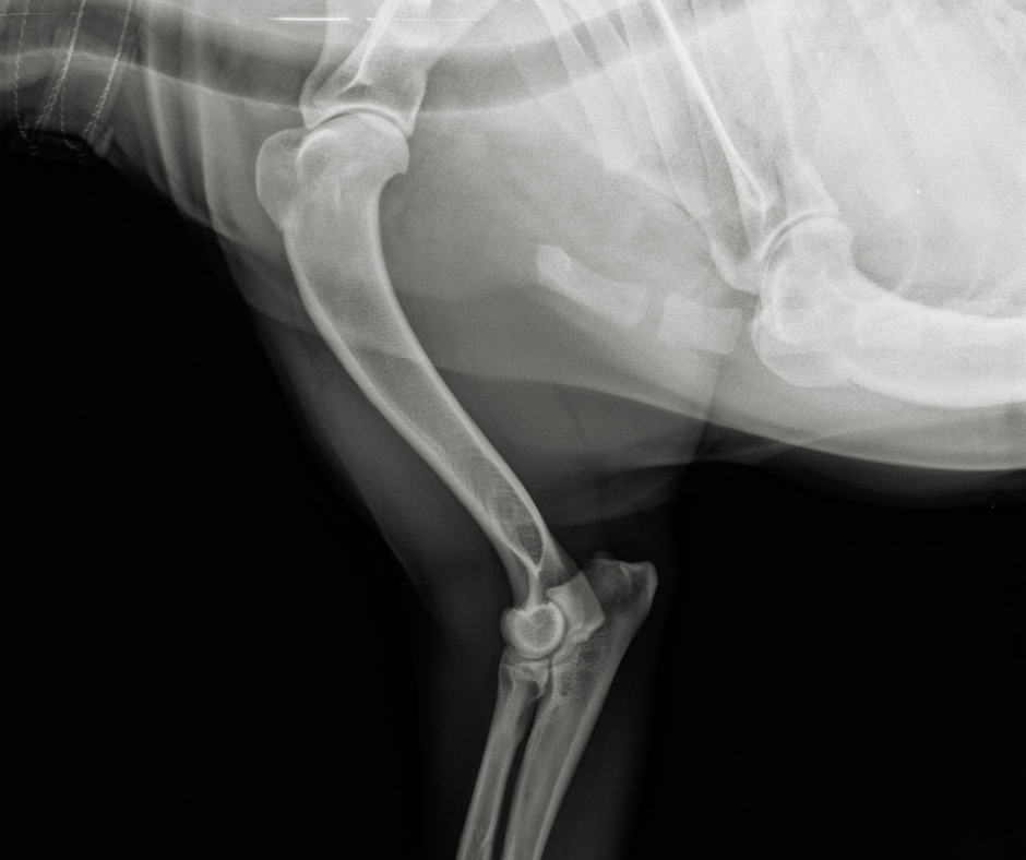
Veterinary Musculoskeletal Diagnostic Ultrasound is a non-invasive imaging tool that allows us to evaluate soft tissue structures such as tendons, ligaments, muscles, and joint capsules in real time. Using high-frequency sound waves, this technology provides detailed images that help identify strains, tears, inflammation, and other subtle injuries that may not be visible on radiographs (X-rays).
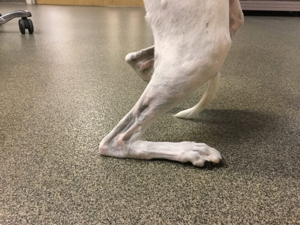
Because ultrasound shows structures dynamically, we can assess tissues both at rest and during movement—giving us valuable insight into how an injury is functioning, not just how it looks. This makes it especially useful for active and sporting dogs, as well as pets with lameness, swelling, or persistent soft tissue pain.
Musculoskeletal ultrasound is painless, and allows for precise diagnosis and targeted treatment planning. It also enables us to monitor healing over time, ensuring your pet’s rehabilitation program is progressing safely and effectively.
Common Ultrasound Questions
-
The most common areas for diagnostic ultrasound include the shoulder, Achilles tendon and iliopsoas muscle, though many others can be imaged as well.
-
Ultrasound can be used to determine the severity of an injury as well as assess the healing process over time. It can impact treatment decisions including what treatment modalities should be performed as well as if injections, bracing, or surgery is needed.
-
Ultrasound appointments are typically scheduled for one hour.
-
Yes, ultrasounds can be scheduled alongside other procedures, typically for ultrasound-guided injections or shockwave therapy.
-
Depending on the area we are imaging, it may be necessary to sedate your pet in order to ensure their comfort and get quality images. Pets are monitored closely and the sedation is reversible which allows them to recover quickly.